9 Aleq Manukyan St. Rooms 202,102 , AUA Business Centre Yerevan, 0070, Armenia
Ալեք Մանուկյան, 9,202,102 սենյակներ, ՀԱՀ բիզնես կենտրոն Երեւան 0070, Հայաստան

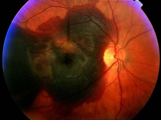
Age Related Macular Degeneration (AMD/ARMD)
Տարիքային մակուլոդիստրոֆիա
Տարիքային մակուլոդիստրոֆիան հիմնականում հանդիպում է 50 տարեկանից բարձր անձանց մոտ և բնորոշվում է ցանցաթաղանթի կենտրոնական հատվածի` մակուլայի փոփոխություններով, որոնք հանգեցնում են կենտրոնական տեսողության վատացմանը:
Տարիքային մակուլոդիստրոֆիան կարող է լինել չոր (90%) կամ թաց (10%): Չոր ձևը կարող է վերածվել թացի: Թաց մակուլոդիստրոֆիան ի տարբերություն չորի կարող է կարճ ժամանակահատվածում հանգեցնել տեսողության զգալի վատացման կամ կորստի: Թաց մակուլոդիստրոֆիայի ժամանակ ցանցաթաղանթի տակ առաջանում են արյունատար անոթներ, որոնք շատ հեշտ վնասվելու հակում ունեն: Այդ անոթների վնասման հետևանքով ցանցաթաղանթի նորմալ կառուցվածքը խախտվում է` հանգեցնելով կենտրոնական տեսողության վատացմանը:
What is AMD?
AMD - is a very common condition, usually associated with advanced age. It is a condition where the retina deteriorates leading to blurry vision, trouble seeing detail and reading, distortion or wavy vision.
Այդ պատճառով մակուլոդիստրոֆիայի վաղ ախտորոշումն ու պարբերաբար ստուգումները չափազանց կարևոր են:
Dry and Wet AMD
There are two forms of AMD – dry (90%) and wet (10%). The dry form precedes the wet, although only a minority of patients will develop the wet form. The typical clinical sign of dry AMD is drusen (small yellowish deposits). Drusen appear under the retina and if, advanced, change the shape of the retina, thus causing wavy and distorted central vision. Drusen progress slowly and if diagnosed in early stages correction in diet, regular eye check ups and some recommendations given by an eye doctor can decrease the risk of AMD progression.
In the wet form of macular degeneration, newly created abnormal blood vessels grow under the center of the retina. These blood vessels leak, bleed and scar the retina, distorting or destroying central vision. Vision distortion usually starts in one eye and may affect the other eye later.
In contrast to the dry type, vision loss may be rapid in the wet type of AMD.
Risk factors for AMD
Ռիսկի գործոնները
- Positive family history
- Smoking
- 50 years old and above
- Diet lacking in vitamins and minerals
- Cardiovascular diseases/Hypertension
- Ծնողների մոտ/բարեկամների մոտ մակուլոդիստրոֆիայի առկայությունը
- Ծխելը. կատարված բազմաթիվ հետազոտություններն ապացուցել են, որ ծխելը բարձրացնում է մակուլոդիստրոֆիայի հավանականությունը 6 անգամ:
- Վիտամիններով և միկրոտարրերով աղքատ սննդակարգը
- Սիրտանոթային հիվանդությունները/Հիպերտանիան
Recommendations to minimize AMD.
Մակուլոդիստրոֆիայի առաջացման հավանականությունը նվազեցնելու համար խորհուրդ է տրվում`
- Quit smoking - smoking increases the chance of AMD 6 times.
- Protect your eyes from sunlight – always wear sunglasses when you are outdoors (the blue light damages the pigment layer of the retina)
- Change your diet – avoid high fat foods, increase leafy vegetables in your diet
- Include eye supplements with antioxidants in your diet
- Have regular eye check ups
- Test your central vision using the Amsler grid test between your routine eye check ups
- Չծխել
- Պաշտպանել աչքերն արևից. դրսում կրել արևային ակնոց կամ լայնեզր գլխարկ
- Պահպանել վիտամիններով, միկրոտարրերով և չհագեցած ճարպաթթուներով հարուստ սննդակարգ (կանաչ և կարմիր բանջարեղեն, ծովային սնունդ)
- Ընդունել վիտամինային հավելումներ
- Պարբերաբար ստուգել աչքերը
- Ստուգումների միջև ընկած ժամանակահատվածում կենտրոնական տեսողությունը տուգել Ամսլերի ցանցով:
Along with regular examinations by an eye doctor, people can evaluate their eyesight for possible symptoms of AMD using a simple home testing device known as the Amsler grid. The Amsler grid, consisting of parallel and perpendicular lines, looks much like a sheet of graph paper. By focusing on a marked spot in the middle of the grid, it is quite easy to detect blurred or distorted vision.
Ամսլերի ցանցը հորիզոնական և ուղղահայաց գծերից կազմված մի ցանց է, որի կենտրոնական հատվածում կա սև կետիկ:

How to use the Amsler Grid Test?
Ինչպես օգտվել Ամսլերի թեստից
- If you wear reading glasses, put them on.
- Hold the Amsler grid at a normal reading distance, approximately 30-35 cm.
- Cover one eye and focus on the black dot in the center
-
Do any of the lines look wavy, blurred or distorted?
- Are there any missing areas or dark areas in the grid?
- Can you see all corners and sides of the grid?
VERY IMPORTANT: Don’t forget to test both eyes.
All lines should be straight, all intersections should form right angles and all the squares should be the same size. Report any irregularity to your eye doctor immediately. You can mark areas of the chart that you’re not seeing properly and bring it with you to your eye exam.
- Եթե Դուք ակնոց եք կրում, ապա թեստը կատարեք ակնոցով:
- Պահեք Ամսլերի ցանցը դեմքից մոտավորապես 30-35 սմ հեռու:
- Ձեռքով փակեք մեկ աչքը և ուշադիր նայե՛ք կենտրոնում գտնվող կետին: Կրկնեք թեստը մյուս աչքի համար:
-
Անմիջապես այցելեք Ձեր ակնաբույժին, եթե`
- ցանցի գծերն ալիքավոր կամ ծռմռված եք տեսնում,
- քառակուսիները չափով կամ ձևով տարբերվում են իրարից,
- ցանցի որևէ հատված բացակայում է կամ պղտոր է երևում: